


EyeSight AI: Retinal Image Analysis Software
Computer vision that helps eye-care hospitals review retinal images, with the AI models and the application around them built by KoderTroop.
Computer Vision for Eye-Care Teams
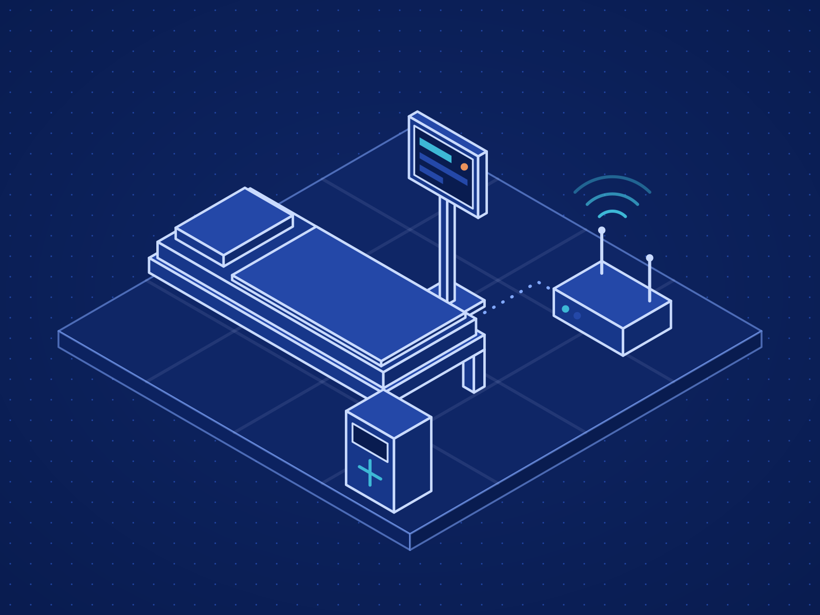
EyeSight AI analyzes retinal images to support the diagnosis of eye diseases in eye-care hospitals. KoderTroop built the software and the AI models. The models run inside an application designed around how a hospital captures, reviews and records images, so their output reaches clinicians where they already work. Every result informs a clinician's assessment, and the decision remains theirs.
How EyeSight AI Works
The screens your team works in each day, and what each one is for.
| Study | Patient | Eyes | Captured | Stage | Clinician | Waiting |
|---|---|---|---|---|---|---|
| ST-2292 | PT-10490 | OS | 09:18 · Camera 1 | Captured | Unassigned | 2 min |
| ST-2291 | PT-10482 | Both | 09:12 · Camera 2 | Analysed | ARA. Rao | 8 min |
| ST-2290 | PT-10477 | OD | 09:05 · Camera 1 | In review | MIM. Iyer | 15 min |
| ST-2289 | PT-10469 | Both | 08:58 · Camera 2 | Recapture | Unassigned | 22 min |
| ST-2287 | PT-10455 | Both | 08:40 · Camera 1 | Analysed | ARA. Rao | 40 min |
| ST-2286 | PT-10448 | OS | 08:31 · Camera 2 | In review | SDS. Das | 49 min |
| ST-2285 | PT-10441 | Both | 08:22 · Camera 1 | Signed off | MIM. Iyer | — |
| ST-2284 | PT-10436 | Both | 08:10 · Camera 2 | Recorded | ARA. Rao | — |
| ST-2283 | PT-10430 | OD | 08:02 · Camera 1 | Recorded | SDS. Das | — |
| Study | Images | Quality | Model | Total | Now |
|---|---|---|---|---|---|
| ST-2292 | 1 | Passed | — | 0.7 s | Analysing |
| ST-2291 | 2 | Passed | 1.8 s | 3.1 s | Review queue |
| ST-2290 | 1 | Passed | 1.7 s | 2.9 s | Review queue |
| ST-2289 | 2 | Recapture | — | 0.9 s | Back to camera room |
| ST-2288 | 2 | Passed | 1.9 s | 3.2 s | Review queue |
| ST-2287 | 2 | Passed | 1.8 s | 3.0 s | Review queue |
| ST-2286 | 1 | Passed | 1.6 s | 2.8 s | Review queue |
| ST-2285 | 2 | Passed | 1.8 s | 3.1 s | Recorded |
R1 confirmed. R2 is a camera artefact. Repeat imaging at the next routine visit and compare with today.
- R1 · small bright spots, superior arcade · confirmed
- R2 · dark spot, temporal to macula · overridden
- R3 · vessel calibre change, inferior arcade · confirmed
Model findings were reviewed by the signing clinician. This report records the clinician’s assessment.
Computer-vision models analyze each retinal image and highlight findings for a clinician to review.
Quoted Per Deployment
EyeSight AI is priced to the scope of each deployment: sites, devices, integrations and the support you need.
Quote for EyeSight AI
Tell us how many sites and devices you operate and which systems it must connect to, and we will return a scoped quote.